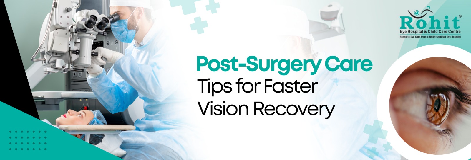
Undergoing eye surgery is a major step toward better vision and improved quality of life. Whether you’ve had cataract surgery or LASIK, the procedure itself is only part of the journey. Proper recovery and aftercare play an equally important role in achieving the best possible visual outcomes.
At Rohit Eye Hospital, we often remind patients that healing doesn’t stop in the operating room. Following the right cataract surgery aftercare and practical LASIK recovery tips can significantly improve comfort, speed up healing, and protect your long-term eye health.
This guide will help you understand what to expect after surgery and how to care for your eyes during the recovery phase.
Understanding the Importance of Post-Surgery Eye Care
Eye surgeries such as cataract removal and LASIK are advanced, safe, and highly successful procedures. However, your eyes need time to heal and adjust after the intervention.
The cornea and surrounding tissues undergo microscopic changes during surgery. Although modern techniques are minimally invasive, your body still needs proper care and rest to recover fully. Neglecting post-operative instructions can delay healing or, in rare cases, lead to complications.
The good news is that recovery is usually smooth when patients follow medical advice carefully. With a little patience and attention, you can return to your normal routine quickly while enjoying clearer vision.
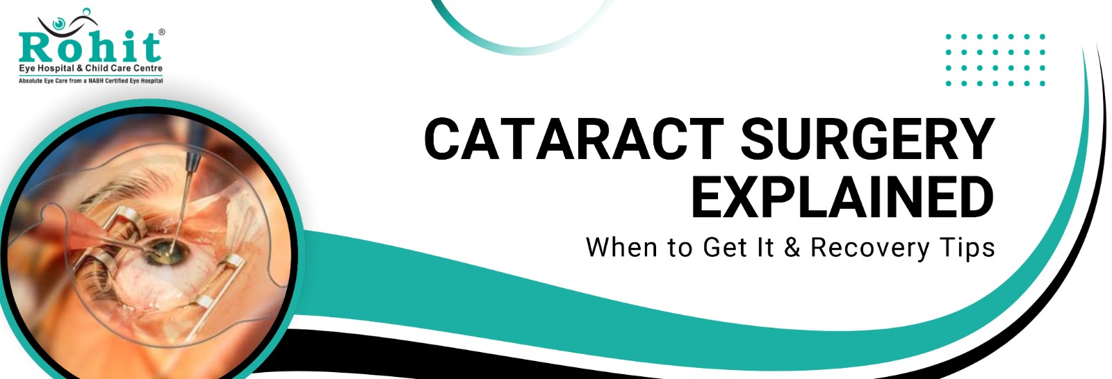
Cataract Surgery Aftercare: Essential Guidelines for Smooth Healing
Cataract surgery is one of the most commonly performed and safest procedures worldwide. Most patients notice improved vision within a few days. Still, complete healing may take a few weeks.
Here are the most important cataract surgery aftercare steps:
1. Use Prescribed Eye Drops Consistently
Your doctor will prescribe antibiotic and anti-inflammatory eye drops. These medications prevent infection, reduce swelling, and support healing.
Never skip doses or stop using drops early unless your doctor advises it. Even if your eye feels normal, internal healing is still in progress.
2. Avoid Rubbing or Pressing the Eye
It is common to feel mild itching or a foreign-body sensation after surgery. Resist the urge to rub your eyes. Rubbing can disturb the healing incision or introduce bacteria.
3. Protect Your Eyes from Dust and Bright Light
Wear protective glasses or sunglasses when stepping outdoors. This helps shield your eyes from dust, pollution, and harsh sunlight, especially in the first week after surgery.
4. Sleep Carefully
Try not to sleep on the side of the operated eye for a few days. Many patients are advised to wear an eye shield at night during the early recovery period.
5. Avoid Heavy Lifting and Strenuous Activity
Activities that increase pressure inside the eye should be avoided for at least a week. This includes bending excessively, lifting heavy objects, or engaging in intense workouts.
6. Maintain Hygiene
While bathing, ensure that soap and water do not enter the eye. Avoid swimming pools, hot tubs, or dusty environments until your doctor confirms it is safe.
Following these simple but critical cataract surgery aftercare steps ensures stable healing and optimal visual clarity.
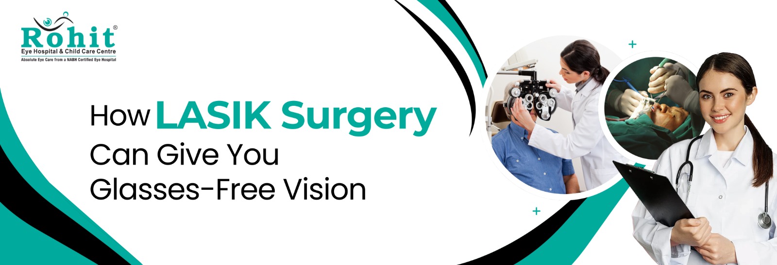
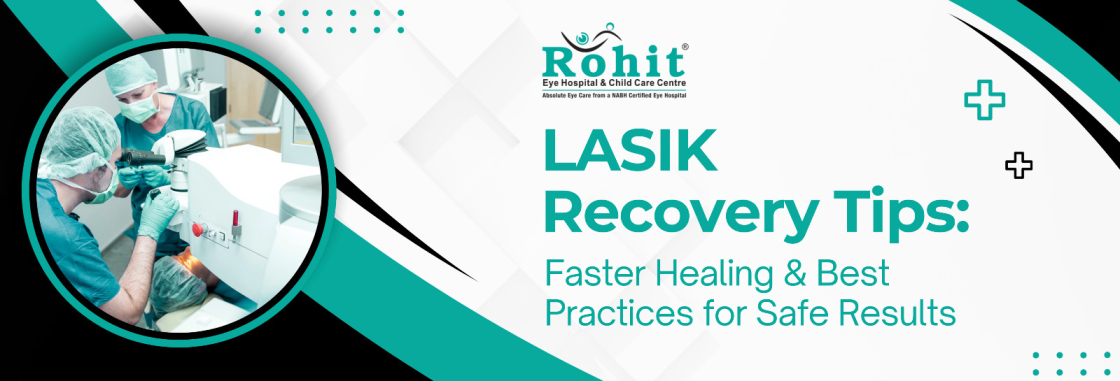
LASIK Recovery Tips: What to Do for Fast and Comfortable Healing
LASIK is a popular refractive procedure designed to correct vision problems like myopia, hyperopia, and astigmatism. The recovery process is typically faster than most people expect, but proper care still matters.
Here are practical LASIK recovery tips to help you heal smoothly:
1. Rest Your Eyes Immediately After Surgery
You may experience mild burning, watering, or sensitivity to light for a few hours after LASIK. Resting with closed eyes helps the corneal flap settle naturally.
2. Avoid Screens Initially
Limit screen time for the first 24 to 48 hours. Extended exposure to mobile phones, laptops, or television can increase dryness and strain.
3. Use Lubricating Drops Regularly
Dryness is common after LASIK. Artificial tears help maintain comfort and promote healing. Even if your eyes feel fine, continue using the drops as advised.
4. Do Not Touch or Rub Your Eyes
The corneal flap created during LASIK needs time to stabilize. Avoid rubbing your eyes for at least a few weeks.
5. Avoid Makeup and Eye Products
Refrain from using eye makeup, creams, or lotions near the eyes during the initial healing period to prevent infection.
6. Skip Swimming and Contact Sports
Activities that expose your eyes to water or physical impact should be avoided for at least two weeks.
These LASIK recovery tips may seem simple, but they make a significant difference in achieving stable and long-lasting results.
What to Expect During Vision Recovery
It is normal for vision to fluctuate slightly during the first few days after surgery. Some patients notice halos, glare, or mild sensitivity to light. These symptoms gradually improve as healing progresses.
For cataract patients, colors may appear brighter and clearer because the cloudy lens has been replaced with a transparent artificial lens. LASIK patients often experience immediate clarity, though fine adjustments in vision may continue for several weeks.
Recovery timelines vary from person to person. Factors such as age, overall eye health, and adherence to post-operative instructions influence healing speed.
Diet and Lifestyle for Faster Eye Healing
Your body heals better when supported with proper nutrition and rest. Consider these additional recovery tips:
- Stay hydrated throughout the day.
- Include vitamin-rich foods such as leafy greens, carrots, citrus fruits, and nuts.
- Get adequate sleep to promote tissue repair.
- Avoid smoking, as it slows down healing.
Healthy habits complement medical care and contribute to long-term eye health.
Warning Signs: When to Contact Your Eye Doctor
While complications are rare, it is important to stay alert. Seek medical advice immediately if you experience:
- Sudden decrease in vision
- Severe or increasing eye pain
- Persistent redness
- Excessive discharge
- Flashes of light or floating spots
Prompt consultation ensures that minor issues are addressed before they become serious.
Follow-Up Appointments Are Crucial
Many patients feel fine within a day or two and assume follow-ups are unnecessary. However, scheduled check-ups allow your ophthalmologist to monitor internal healing, eye pressure, and visual stability.
Attending follow-up visits is an essential part of cataract surgery aftercare and LASIK recovery tips. These appointments ensure that your vision is progressing as expected.
Emotional and Psychological Adjustment After Vision Surgery
Interestingly, some patients experience an emotional adjustment period after achieving clearer vision. If you have worn glasses for many years, adapting to glasses-free clarity can feel surprising at first.
Give yourself time to adjust. Your brain needs a short period to adapt to improved visual input. This phase is completely normal and temporary.
Long-Term Eye Care After Surgery
Even after complete recovery, maintaining regular eye check-ups is important. Eye health is dynamic and can change with age.
Here are long-term recommendations:
- Continue routine annual eye exams.
- Wear UV-protective sunglasses outdoors.
- Manage systemic conditions like diabetes or hypertension.
- Maintain a balanced lifestyle to protect visual health.
Surgery corrects existing issues, but preventive care protects your vision for the future.
Why Proper Aftercare Determines Surgical Success
Modern eye surgeries boast success rates above 95 percent. However, patient participation during recovery is what transforms a successful procedure into a truly life-changing outcome.
Following cataract surgery aftercare instructions prevents infection, reduces inflammation, and stabilizes the implanted lens. Similarly, adhering to LASIK recovery tips ensures proper corneal healing and long-term refractive stability.
In short, surgery provides the foundation, and aftercare builds the results.
Trusted Post-Operative Support at Rohit Eye Hospital
At Rohit Eye Hospital, patient care extends beyond the operation theatre. Our team provides detailed recovery guidance, personalized follow-up plans, and continuous support throughout the healing journey.
We understand that every patient is different. Age, lifestyle, and eye condition influence recovery needs. That’s why post-surgical care plans are tailored individually.
From detailed medication instructions to emergency support, we ensure that your recovery remains safe, comfortable, and predictable.
Conclusion
Vision surgery is a powerful step toward clearer sight and greater independence. Whether you have undergone cataract surgery or LASIK, the healing phase deserves careful attention.
By following essential cataract surgery aftercare steps and practical LASIK recovery tips, you can accelerate healing, reduce discomfort, and protect your long-term visual outcomes.
Clear vision is not just about the procedure itself. It is about the care you take afterward. With proper guidance, patience, and support from experienced professionals, your eyes can heal smoothly and deliver the clarity you’ve been waiting for.
If you are planning eye surgery or have recently undergone one, consult the specialists at Rohit Eye Hospital for expert guidance and comprehensive post-operative care. Your journey to better vision deserves nothing less than the highest standard of attention and expertise.